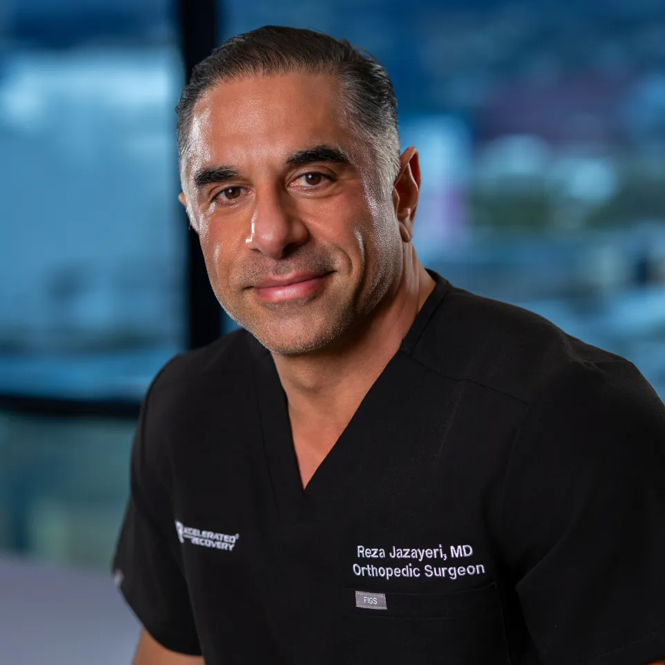
Rotator cuff tears can make everyday movements feel difficult and painful. If you live in the Greater Los Angeles area and shoulder pain is holding you back, you need clear, practical information about your options. Dr. Reza Jazayeri is a double board-certified orthopedic surgeon specializing in sports medicine, with clinical expertise in shoulder, knee, and elbow care.
He completed his sports medicine fellowship at the renowned Kerlan-Jobe Orthopaedic Clinic in Los Angeles. Dr. Jaz sees patients at his Beverly Hills, Westlake Village, and Orange offices. His practice explores non-surgical paths first and uses minimally invasive techniques when surgery is needed. This page walks you through what rotator cuff tears are, how they are treated, and what to expect during recovery.
Understanding Your Diagnosis and Treatment Options
A clinical overview of partial and full-thickness rotator cuff pathology, the spectrum of non-operative and surgical management, and the evidence supporting orthobiologic and peptide-based treatment strategies.
Anatomy of the Rotator Cuff
The rotator cuff is a group of four muscles and their tendons that surround and stabilize the glenohumeral (shoulder) joint. Together, they allow the arm to lift and rotate while keeping the humeral head centered within the shoulder socket.
The Four Muscles
- Supraspinatus — initiates shoulder abduction; the most commonly injured tendon
- Infraspinatus — controls external rotation
- Teres Minor — assists external rotation and inferior stabilization
- Subscapularis — primary internal rotator and anterior stabilizer
Why Tendons Are Slow to Heal
Tendons — particularly the supraspinatus near its bony insertion — have inherently poor blood supply. This hypovascular environment limits oxygen, nutrient, and repair cell delivery needed to mount an effective healing response [1]. In many patients, tears occur on a background of underlying degenerative tendinopathy — disorganized collagen, reduced cell activity, and accumulated damage that further impairs healing.

Classifying Rotator Cuff Tears
Rotator cuff tears exist on a spectrum of severity. Where your tear falls on this continuum directly determines the most appropriate treatment strategy. Not all tears require surgery.
| CLASSIFICATION | DEPTH | SYMPTOMS | PRIMARY APPROACH | SURGICAL THRESHOLD |
|---|---|---|---|---|
| Low-Grade Partial | <25% thickness(Ellman Grade I) | Mild aching, activity pain, preserved strength | Physical therapy, activity modification, PRP→ Non-operative | Rarely indicated |
| High-Grade Partial | 25–75% thickness(Ellman Grade II–III) | Significant pain, night pain, weakness with resisted elevation | Physical therapy + PRP ± BMAC, metabolic optimization→ Non-operative preferred | After ≥3–6 months of conservative care failure. 2023 study: ASES scores 53.2 → 92.9 at 2-year follow-up with PRP alone [16] |
| Full-Thickness | 100% thickness(Complete disruption) | Significant weakness, difficulty lifting arm, pain with ADLs | Physical therapy as bridge; small tears may respond to PRP→ Surgery often indicated | Recommended for active patients with functional limitation after conservative management [3] |
The Treatment Continuum
Management follows a stepwise approach guided by tear severity, symptoms, and each patient’s individual biology.
Step 01 — Physical Therapy & Activity Modification
Targeted rehabilitation addresses rotator cuff strength, scapular mechanics, and neuromuscular control. Physical therapy is the cornerstone of non-operative management and a prerequisite to any surgical intervention.
Step 02 — Orthobiologic Injection Therapy
Platelet-rich plasma (PRP) and bone marrow aspirate concentrate (BMAC) are evidence-supported, in-office treatments that use the body’s own biological resources to stimulate tissue repair and healing.
Step 03 — Surgical Repair (When Indicated)
Arthroscopic rotator cuff repair is reserved for full-thickness tears with functional limitation in patients who have not responded to conservative care.
Orthobiologic Treatment Options
Orthobiologics are substances derived from the patient’s own body — termed autologous — used to enhance the biological healing of musculoskeletal tissue. Because they come from the patient, there is no risk of immune rejection or cross-contamination.
Platelet-Rich Plasma (PRP)
PRP is a concentration of the patient’s own platelets prepared from a peripheral blood draw. Upon delivery to the injury site, activated platelets release platelet-derived growth factor (PDGF), transforming growth factor-β (TGF-β), vascular endothelial growth factor (VEGF), and insulin-like growth factor-1 (IGF-1) — collectively driving cell recruitment, collagen synthesis, angiogenesis, and matrix remodeling at the tear site [6].
- Tenocyte Stimulation: Promotes proliferation of tendon cells and collagen production
- Angiogenesis: VEGF drives new blood vessel formation, improving nutrient delivery to the hypovascular tendon
- Inflammatory Modulation: Shifts the local environment from chronic destructive inflammation toward active tissue repair
- Collagen Remodeling: TGF-β promotes type I collagen synthesis, improving tendon structural quality
Bone Marrow Aspirate Concentrate (BMAC)
BMAC is obtained by aspirating bone marrow — typically from the posterior iliac crest — and concentrating it through centrifugation. BMAC is one of the richest autologous sources of mesenchymal stem cells (MSCs) available in clinical practice. MSCs are multipotent cells capable of differentiating into tendon, cartilage, and bone lineages. Beyond direct regenerative potential, MSCs exert powerful paracrine effects — secreting bioactive molecules that modulate local inflammation and create a permissive environment for repair [7] [8].
- Rich MSC Content: Bone marrow is the premier autologous MSC source — capable of differentiating toward tendon, cartilage, and bone lineages
- Paracrine Signaling: MSCs release HGF, IGF-1, VEGF, and IL-10, suppressing inflammation and recruiting native repair cells
- Immunomodulation: MSCs convert a destructive chronic inflammatory state into a pro-healing environment
- Structural Contribution: MSC-derived tenocytes contribute directly to collagen matrix deposition and tendon remodeling

PRP Preparation: Not All PRP Is Equal
The clinical outcome of PRP therapy depends critically on preparation quality. Multiple variables determine platelet concentration, growth factor yield, and biological activity.
Step 01 — Blood Collection
Adequate blood volume — typically 30–60 mL — is required to yield a sufficient platelet concentrate. Excessive mechanical shear during draw can activate platelets prematurely, depleting the growth factor payload before injection [9].
Step 02 — Centrifugation
A validated two-spin protocol concentrates platelets to a therapeutic level — typically 3–5× baseline platelet count. The g-force and duration of each spin are calibrated to maximize yield without activating or damaging the cells [10].
Step 03 — Formulation: Leukocyte Content
The appropriate leukocyte (white blood cell) content depends on the tissue being treated and the nature of the pathology — this is not a one-size-fits-all decision [11].
- Leukocyte-Rich PRP (LR-PRP): Preferred for tendon pathology and partial tears; more robust initial inflammatory stimulus for chronically degenerate tendon tissue; not preferred for intra-articular injections
- Leukocyte-Poor PRP (LP-PRP): Preferred for intra-articular (joint space) injections; reduced MMP activity protective of cartilage and synovium; lower risk of post-injection inflammatory flare.
Ultrasound-Guided Delivery
All orthobiologic injections are performed under real-time musculoskeletal ultrasound guidance. Landmark-guided shoulder injections miss the target in a significant proportion of cases; ultrasound allows direct visualization of needle tip position, confirmation of injectate spread within the target tissue, and protection of adjacent neurovascular structures [12]. The full procedure — blood draw, centrifugation, and injection — is completed in the outpatient office setting without anesthesia.
Metabolic Optimization: Preparing the Biology
The biological environment in which healing occurs is as important as the intervention itself. Dr. Jazayeri evaluates and corrects modifiable variables prior to every procedure.
- Vitamin D Status: Critical for musculoskeletal repair, immune regulation, and platelet function; deficiency is corrected before treatment
- Hormonal Profile: Sex hormones regulate collagen synthesis and regenerative capacity; suboptimal status is addressed where clinically appropriate
- Inflammatory Markers: Chronic systemic inflammation — reflected in hsCRP and cytokines — creates an unfavorable environment for repair; reducing pro-inflammatory states improves treatment response
- Metabolic Screening: Glucose homeostasis, insulin sensitivity, and thyroid function are assessed; insulin resistance is associated with inferior tendon quality and is addressed where modifiable
- Nutritional Supplementation: Collagen precursors (vitamin C, proline, glycine), omega-3 fatty acids, and key micronutrients are provided before and after the procedure to support matrix synthesis
- Targeted Physical Therapy: Orthobiologic injection is paired with a structured rehabilitation protocol designed to mechanically load the healing tendon at appropriate intervals, driving collagen fiber alignment
Peptide Therapeutics: An Adjunct to Orthobiologic Care
For selected patients, Dr. Jazayeri may discuss the potential role of therapeutic peptides as an adjunct to orthobiologic treatment. Peptides are short chains of amino acids that function as precision signaling molecules — biological messengers that communicate with cells to upregulate specific repair pathways, promote collagen synthesis, support angiogenesis, and modulate the inflammatory environment.
BPC-157 — Body Protective Compound-157
A synthetic pentadecapeptide with demonstrated activity in preclinical tendon healing models. BPC-157 promotes angiogenesis through the VEGF pathway, stimulates fibroblast migration and proliferation, and upregulates growth hormone receptors on tendon cells — potentially enhancing their responsiveness to growth factors delivered via PRP or BMAC [22] [23] [24]. Its pro-angiogenic mechanism is particularly relevant in the hypovascular tendon critical zone where healing most commonly stalls.
TB-500 — Thymosin Beta-4 Fragment
A synthetic 43-amino-acid peptide derived from thymosin beta-4, an endogenous protein that regulates actin dynamics and cellular motility. TB-500 enhances migration of repair cells into damaged tendon tissue, promotes new blood vessel formation, modulates pro-inflammatory cytokines, and has been shown to mobilize endogenous stem cells from bone marrow — complementing exogenous BMAC delivery [25].
GHK-Cu — Copper Tripeptide
A naturally occurring tripeptide-copper complex found in human plasma that declines substantially with age. GHK-Cu acts as a gene-level signaling molecule, stimulating fibroblast production of collagen, elastin, and glycosaminoglycans — the structural components of tendon matrix. It also modulates the balance between collagen-degrading enzymes and their inhibitors, promoting organized remodeling rather than uncontrolled breakdown of tendon tissue.
Growth Hormone Secretagogues
Peptides such as CJC-1295 and Ipamorelin stimulate physiological, pulsatile growth hormone release from the pituitary, elevating IGF-1 — a critical mediator of musculoskeletal repair and collagen synthesis. GH and IGF-1 decline with age; restoring physiological signaling creates a systemic anabolic environment that can meaningfully improve the body’s capacity to respond to local orthobiologic interventions.

The Comprehensive Healing Framework
Successful non-operative healing of a rotator cuff tear — particularly in high-grade partial tears — requires coordinating all relevant biological layers simultaneously. Dr. Jazayeri designs an individualized protocol that addresses each component below.
-
Ultrasound-guided PRP and/or BMAC delivers concentrated autologous growth factors and mesenchymal stem cells directly to the tear, initiating and sustaining the healing cascade where native blood supply is inadequate.
-
Peptides promote angiogenesis, mobilize repair cells, upregulate growth factor receptors, and create a systemic anabolic environment through optimized GH/IGF-1 signaling — amplifying the effects of local orthobiologic therapy.
-
Growth factor signals can only drive collagen synthesis if raw materials are present. Targeted supplementation provides collagen precursors, omega-3 fatty acids, and micronutrients for tendon matrix repair — before and through remodeling.
-
Vitamin D deficiency, hormonal insufficiency, insulin resistance, and chronic inflammation each impair healing biology. Correcting these variables ensures the systemic environment actively supports the regenerative interventions.
-
Tendons require calibrated mechanical loading — mechanotransduction — to organize newly synthesized collagen into aligned, load-bearing structures. A tailored rehabilitation protocol provides the mechanical stimulus to convert biological repair into durable tendon tissue.
When Surgery Is Indicated
For patients with full-thickness rotator cuff tears who experience meaningful pain, weakness, and limitation of activities — and who have not responded to structured conservative management — arthroscopic repair represents the standard of care. The procedure reattaches the torn tendon to its bony footprint under arthroscopic visualization using suture anchors, and outcomes in appropriately selected patients are generally favorable [3].
The probability of successful biological healing with surgical reattachment is substantially higher in complete tears than in partial tears. This supports the preferential use of non-operative and regenerative strategies for partial tear populations, reserving surgery for those where clinical evidence most strongly favors reattachment [4].
Criteria for Surgical Consideration
- Full-thickness tear confirmed on MRI or high-resolution ultrasound
- Significant pain impacting quality of life or function
- Weakness with resisted abduction or external rotation
- Failure of 6–12 weeks of structured physical therapy
- Functional limitation affecting daily activities or occupation
- Appropriate tissue quality for successful repair
Meet Dr. Reza Jazayeri
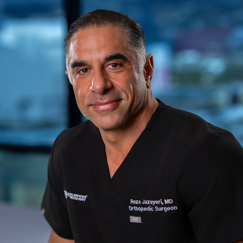
Dr. Reza Jazayeri specializes in sports medicine with expertise in shoulder, knee, and elbow injuries. A Southern California native, Dr. Jazayeri combines an engineering background with elite surgical training, completing his orthopedic surgery residency at the nationally ranked NYU Hospital for Joint Diseases and a sports medicine fellowship at the renowned Kerlan-Jobe Orthopaedic Clinic.
After more than 15 years caring for patients within the Southern California Permanente Medical Group, Dr. Jazayeri joined DOCS Health to expand a practice focused on personalized orthopedic and sports medicine care and surgical excellence. As Director of Longevity at DOCS Health, he leads the development and implementation of optimization protocols and regenerative medicine strategies designed to improve long-term musculoskeletal health and extend healthspan.
His published work includes a 2026 coauthored review of nutritional optimization in spine surgery in the Journal of the American Academy of Orthopaedic Surgeons. View Dr. Jazayeri’s research publications and training.
Dr. Jazayeri is an incredibly skilled surgeon with great communicative skills. He came to me very highly referred. At my initial appointment, I quickly found him to be very knowledgeable, compassionate, and dedicated to his patients’ well-being. I immediately knew I had chosen the correct surgeon to do my should replacement. He took the time to answer all my questions, while at the same time explaining AND showing me on x-rays exactly what was wrong with my shoulder. I was impressed by his commitment to going the extra mile, not only in making sure I understood the upcoming surgery, but also in his confidence in knowing it would be successful. I am now 9 weeks post op, and have had almost zero pain since the procedure. Dr. Jazayeri continues to personally monitor my rehab progress via the “My Health Track” app and timely check ups. I am confident in my continued shoulder healing progress and know I made the correct choice in surgeons. I recommend Dr. Jazayeri very highly and am confident anyone who chooses him for their procedure, will also know they made the right choice in surgeons.
Schedule a Consultation with Dr. Jazayeri
If you have been diagnosed with a rotator cuff tear and would like to explore non-operative regenerative treatment options — including ultrasound-guided PRP, BMAC, or peptide therapy — we invite you to contact our Beverly Hills, Westlake Village, or Orange office for a comprehensive evaluation.
Medically reviewed by Reza Jazayeri, M.D. on